Professor Profile: Associate Professor Deanne Thompson
What is your current role?
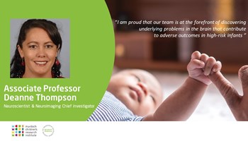
I am a senior neuroscientist and lead the neuroimaging team within Murdoch Children’s Research Institute’s (MCRI) Victorian Infant Brain Studies (VIBeS) group. I am the neuroimaging chief investigator in the CRE and co-chair the research subcommittee with Associate Professor Brett Manley.
How long have you been working in this area? What first interested you and why have you pursued this area of research?
I have been working in the VIBeS team since 2003, when we were still scanning our very first preterm cohort of infants!
Before joining the VIBeS team, I majored in botany, microbiology and genetics at university, and had started a project which involved genetic engineering cotton plants to be resistant to fungus. I soon realised that career was not for me, had a change of heart, and decided to do something to help people more directly! I started searching for research assistant positions in the field of medical research and was attracted to a position within the VIBeS team since I liked the idea helping improve the lives of babies who have a challenging start to life. It was here I found my niche in neuroimaging and continued to do more studies in neuroscience and a PhD.
I am proud that our team has established an international reputation in the field. We are at the forefront of discovering underlying problems in the brain that contribute to adverse outcomes in high-risk infants, especially those born very early. We have also made a major contribution to mapping how the brain develops over time, with our unique cohort of children who have kept returning for follow-up MRI scans since their birth, and are soon to be 20 years old.
How is neuroimaging in particular advanced our understanding of the cohort you are studying?
Our neuroimaging studies have provided crucial information about alterations to the development of the preterm brain, revealing what early life factors contribute to these alterations, and showing how these alterations relate to later outcomes. We have used neuroimaging in multiple randomised controlled trials and interventions to determine how the brain is affected by therapies aimed at improving outcomes of high-risk infants, such as caffeine, steroids, DHA fatty acid and cognitive training.
These trials have led to changes in clinical practice, and neuroimaging findings played a part in this. Along with other international neuroimaging research, our research has led to brain MRI becoming standard of care for very preterm infants in many centres around the world.
Who are your main collaborators?
The CRE’s neuroimaging group is located within the Developmental Imaging team at MCRI, and so we are closely amalgamated with researchers who provide technical expertise for us to successfully apply neuroimaging to our clinical research questions. We have a large network of other collaborators both locally (University of Melbourne; Monash University, including Monash Biomedical Imaging; LaTrobe University; Deakin University; and the Royal Women’s Hospital) and all over the world (Harvard University, including the Computation Radiology Laboratory; Boston Children’s Hospital; Washington University and St Louis Children’s Hospital; Image Sciences Institute, Utrecht, The Netherlands; University of Montreal and University of Calgary in Canada, King’s College London, and others).
What are you now researching?
We are preparing to begin a 20 year follow up of the cohort from which the above findings came. Now that we have finished investigating how the very preterm brain develops, we would like to continue to see how it ages during adulthood.
The neuroimaging team has been very active in creating neuroimaging software tools specific for infants, as these were not available when we began analysing infant MRI scans. We are applying these software tools to other high-risk infant cohorts to determine the effects of adverse early life events, treatments, and interventions on the brain. This technology has also received widespread uptake and is facilitating much more infant brain research across the world than was previously possible. We are also currently working on making these technological advancements available to clinicians, so that when any infant is scanned with an MRI, the doctor will have an instant detailed report on the health of the infant’s brain. This will help them know how to recommend interventions to the family that might be helpful for improving the babies brain development, and hence outcomes.
You can watch Dee talking about her latest publication on tracking regional brain growth in babies born preterm and term at 13 years of age


